


News & Events
What Is Fundus Imaging? A Complete Guide for Tailored to Practice Needs
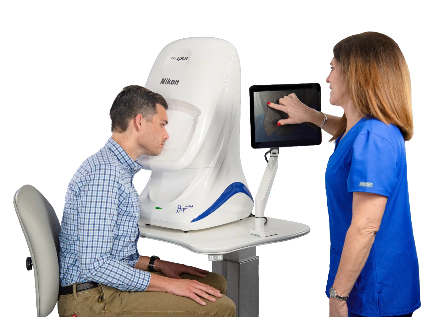
Diabetic eye screening in Singapore runs on retinal photography. But the camera that suits a busy polyclinic or screening center is rarely the one that suits a primary optometrist practice or a specialized retinal clinic.
The gap between these user groups comes down to workflow priorities, delegation level, diagnostic requirements, and patient throughput. Getting the choice wrong results in equipment that either slows down clinic flow or lacks the clinical depth required for advanced pathology management.
This comprehensive guide details what fundus imaging is, how standard and advanced technologies operate, and how to select the right device tailored to your specific clinical tier.
Need a retinal imaging system for your practice?
Browse Vitop’s retinal imaging systems for optometry, ophthalmology and hospital practice or request a quotation
What is Fundus Imaging?
Fundus imaging is a non-invasive diagnostic technique that captures high-resolution photographs of the back of the eye which include the retina, optic disc, macula, and major retinal vessel arcades. It enables eye care practitioners to document baseline structural status, monitor changes over time, and screen for sight-threatening conditions such as diabetic retinopathy (DR), diabetic macular edema (DME), glaucoma, age-related macular degeneration (AMD), and vascular occlusions.
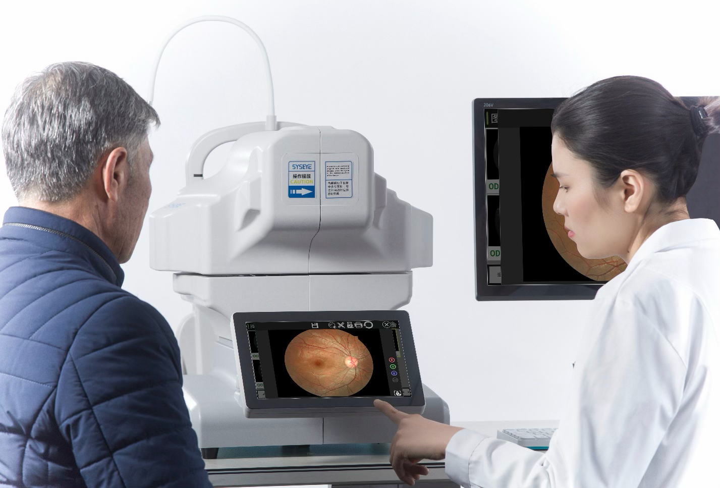
How Fundus Imaging Works
A standard fundus camera functions as a low-power microscope linked to a high-resolution optical sensor. Light enters through the pupil and reflects off the retina along a separate optical path to prevent corneal and lens glare.
- Mydriatic Imaging: Pharmacological dilation expands the pupil, allowing high-resolution visualization into the peripheral retina.
- Non-mydriatic Imaging: Uses infrared light for patient alignment and target fixation, followed by a brief optical flash. Skipping pupil dilation avoids patient wait times, making it the standard choice for screening programs.
Macula & Optic Disc-Centered Two-Field Protocol
Standard 45° to 50° fundus cameras rely on internal target fixations to capture two critical regions per eye:
- Macula-Centered Image: The patient fixates directly forward. Centers the fovea and macula to evaluate central vision changes, microaneurysms, hard exudates, and signs of DME.
- Optic Disc-Centered Image: The internal fixation target shifts, prompting the eye to turn. Centers the optic nerve head to assess cup-to-disc ratios (glaucoma screening), optic disc swelling (papilledema), and nasal retinal vessel architecture.
Fundus Camera Solutions Across 3 Major User Segments
| User Segment | Primary Clinical Focus | Key Device Requirements | Recommended Camera Tier |
|---|---|---|---|
| 1. Polyclinics & Screening Centers | High-volume screening (e.g., SiDRP), rapid triage, automated operation. | Full automation, voice guidance, small pupil capture (≤2.8–3.0mm), DICOM 3.0, AI cloud integration. | Portable Automated (SYSEYE Kestrel 300) / Standard Automated Desktop (SYSEYE Kestrel 3100m) |
| 2. Primary Care Optometrists | Baseline documentation, comprehensive primary eye exams, patient education, in-house referrals. | High-resolution color imaging, automatic alignment, wide stitched fields (135°), fast workflow. | Automated tabletop non-mydriatic camera with montage/stitching capabilities |
| 3. Ophthalmologists & Retinal Specialists | Peripheral retinal pathology, disease staging, surgical management, complex vascular conditions. | 200° single-capture Ultra-Widefield (UWF™), multimodal (FA, FAF, RGB color), OCT integration. | Optos Ultra-Widefield Systems (Daytona, California, Silverstone RGB) |
Segment 1: Polyclinics & High-Volume Screening Programs (SiDRP)
In Singapore, public polyclinics and community screening hubs operate under the Singapore Integrated Diabetic Retinopathy Programme (SiDRP), capturing macula and optic disc-centered photos across over 100,000 diabetic patients annually.
- Operational Needs: Fast image acquisition, minimal dependence on operator skill, non-mydriatic operation, and immediate integration with centralized telemedicine reading centers.
- Portable Automated Advantage (SYSEYE Kestrel 300): Weighing 3.5 kg / 3.6 kg with a portable briefcase setup, the Kestrel 300 features dual-camera autofocus, automatic exposure, built-in voice guidance, and self-service capabilities. Patients complete a binocular examination in under 30 seconds.
- AI Cloud Integration: Captured images transfer directly via DICOM 3.0 / Wi-Fi to AI analysis engines. Deep learning algorithms automatically analyze photographs for microaneurysms and hemorrhages, triaging normal cases automatically and reducing specialist grading workloads by up to 50%.

Segment 2: Primary Care Optometrists
Optometry practices rely on fundus photography to establish structural baselines, document longitudinal changes, build patient compliance, and decide when to initiate specialist referrals.
- Operational Needs: Consistent image quality, easy-to-use operator interfaces, patient communication tools, and multi-field coverage.
- Automated Desktop & Mosaic Solutions: Desktop automated units (such as the SYSEYE Kestrel 3100m) capture precise 50° single fields and stitch up to 9 peripheral shots into a 135° composite widefield image. This provides primary practices with extended peripheral coverage without requiring ultra-widefield laser hardware.
- Patient Education & Engagement: Displaying clear, high-resolution retinal images on-screen helps patients visualize asymptomatic conditions (e.g., early DR or glaucoma disc cupping), improving adherence to recall schedules and medical advice.
Segment 3: Ophthalmologists & Retinal Specialists
Specialist practices managing vitreoretinal disease, severe diabetic retinopathy, retinal detachments, and uveitis require immediate visualization across the far peripheral retina.
- Operational Needs: Single-capture widefield coverage, peripheral lesion detection, multimodal imaging (fluorescein angiography, autofluorescence), and rapid clinic throughput.
- Optos Ultra-Widefield (UWF™) 200° Technology: Optos devices capture a 200° single-capture view (~82% of the retina) through an undilated pupil in under a second.
- Clinical & Efficiency Benefits for Specialists:
- ETDRS Equivalency & Enhanced DR Staging: Optos images show 97% agreement with standard ETDRS 7-field photography within one severity level, while identifying nearly 2-fold more diabetic retinopathy lesions.
- Predominantly Peripheral Lesions (PPL): Up to 50% of diabetic eyes exhibit PPL outside standard fields, increasing DR progression risk in up to 41% of cases and acting as biomarkers for systemic cardiovascular complications.
- Color Modalities (rg vs. rgb): Options include red-green laser scanning (optomap color rg) and red-green-blue (optomap color rgb) for enhanced chorioretinal visualization.
- Clinic Flow & ROI: Centralizing an Optos device reduces total patient visit duration by up to 33% (28 minutes shorter per visit), with clinics reporting complete device payback in under 1 year.
Key Selection Criteria When Choosing Your Fundus Camera

- Field of View: Select based on clinical focus: 45°–50° standard for screening/primary care, 135° stitched for expanded primary documentation, or 200° Optos UWF for retinal specialty care.
- Minimum Pupil Requirement: Opt for devices operating through small pupils (≤2.8mm to 3.0mm) to accommodate elderly or non-dilated screening populations.
- Deployment Style: Portable/handheld units (SYSEYE Kestrel 300) for outreach and mobile health, versus tabletop desktop units or dedicated UWF devices for fixed clinic rooms.
- Software & Connectivity: Native DICOM 3.0 compliance, cloud backup, and direct compatibility with AI grading software.
- Local Technical Support: Ensure comprehensive local vendor support for installation, calibration, preventive maintenance, and quick turnarounds on technical service.
Get Your Fundus Camera From Vitop Singapore
Vitop has supplied and serviced ophthalmic equipment in Singapore and Malaysia since 2013, working with opticians, optometrists, hospitals, eye care clinics and optical laboratories.
Our portfolio of fundus cameras covers all three user tiers, including mobile screening units like the SYSEYE Kestrel 300, automated desktop systems like the SYSEYE Kestrel 3100m and advanced Optos Ultra-Widefield systems.
We handle installation, calibration, preventive maintenance, and operator training locally.
Request a quotation today to match the ideal fundus camera system to your practice workflow and clinical requirements.