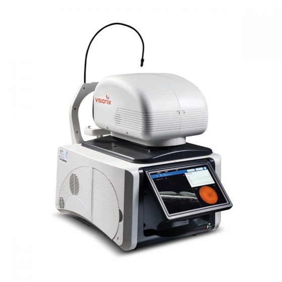
Optovue Autofusion – Automated Dual-Modality OCT & Fundus Camera
The Optovue Autofusion by Visionix combines high-resolution, non-contact Optical Coherence Tomography (OCT) with a 12 MP non-mydriatic colour fundus camera in one fully automated, all-in-one system. With a built-in operating system and integrated touch screen, it needs no external PC – one connection and you are ready to image. Designed for delegation, it lets any team member capture posterior and anterior segment exams with a single tap.
Key Features
- 80,000 A-scans/s OCT – 840 nm light source with < 6 µm axial (in-depth) resolution.
- 12 MP true-colour fundus camera – 45° field of view, non-mydriatic.
- One-tap automation – automatic 3D tracking, alignment, focus and capture.
- Macula + optic nerve in one scan – a single 3D Wide scan delivers both analyses.
- 10 scan modes covering posterior and anterior segment.
- Anterior segment module (CAM, optional) – automated pachymetry and angle measurement.
- Follow-up / progression reports – RNFL trend and significance analysis for glaucoma management.
- 10.1" tilting touch screen, motorised height-adjustable chinrest.
- DICOM connectivity (optional), JPEG/PNG export, USB, LAN and HDMI.
Ophthalmic Imaging, Simplified
- Intuitive use – easy for both experienced and new operators.
- Total autonomy – integrated computer reduces clutter and technical risk.
- Simple installation – one connection and an integrated display.
- Fully automatic process – a single button starts the examination.
- Automatic eye tracking – for accurate, repeatable images.
- Delegation ready – helps clinics meet growing patient demand.
Advanced Imaging Tools
- En Face Viewer – OCT/fundus overlay on any designated area.
- 3D Viewer – reconstructs retinal tissue in 3D for inspection from any angle and scale.
- Anterior Segment Measurement – from precise pachymetry maps to detailed angle evaluation.
Comprehensive Reports
- 3D Wide (OD / OU) – optic disc and macula analysis from one scan.
- 3D Optic Disc – RNFL thickness, disc topography, ONH parameters (cup/disc ratio, rim area, cup volume).
- 3D Macula – ETDRS thickness maps and GCC analysis.
- Line, Wide Line, 5-Line Cross and Radial reports.
- OCT Follow-Up Report – change in RNFL thickness, RNFL trend analysis, RNFL significance map and disc topography over time.
- Anterior – corneal pachymetry map (OD/OS/OU) and anterior chamber angle report.
Reports are pre-formatted and can be exported, printed or shared easily.
How It Works
Macula & optic disc OCT: 1) Select a measurement mode → 2) Tap the centre of the pupil – the system aligns automatically → 3) Press START – the system tracks and completes the scan → 4) Results appear instantly on the preview page.
Cornea (with CAM module): 1) Insert the forehead and cornea anterior module → 2) Select Pachymetry or Angle → 3) Tap the pupil centre or angle → 4) Press START → 5) Results appear instantly.
Technical Specifications
| OCT Scanning |
|---|
| Light source | 840 nm |
| Scan speed | Max. 80,000 A-scans/s |
| In-depth resolution | < 6 µm |
| Scan modes | 3D: 3D Optic Disc, 3D Macula, 3D Wide
Line: Line, Wide Line, 5-Line Cross, Radial
Anterior: Radial Pachymetry, Angle Measurement |
| Scan range | Line & 3D: 6 × 6 mm; Wide Line: up to 12 mm |
| Minimum pupil (OCT) | Ø 2.5 mm |
| Fixation | Internal: 15 points (green LED); External: 1 point (amber LED) |
| Fundus Camera |
|---|
| Image mode | Colour, 12 MP |
| Field of view | 45° |
| Image resolution | Centre 60 lines/mm; Middle 40 lines/mm; Periphery 25 lines/mm |
| Pixel pitch on fundus | 4.3 µm |
| Illumination | Capture: white LED; Alignment: NIR LED |
| Diopter compensation | -15 D to +10 D (no lens); -30 D to -10 D and +5 D to +30 D (internal compensation lens) |
| Minimum pupil (fundus) | Ø 3.8 mm |
| Focus | Auto / Manual (split-image) |
| General |
|---|
| Alignment | Automatic 3D tracking; Manual |
| Chinrest | Motorised |
| Display | 10.1" LCD touch screen, 1280 × 800, tilting |
| Operation range | Front/Back 65 mm; Left/Right 100 mm; Up/Down 30 mm |
| I/O ports | LAN (RJ45), USB 3.0, USB 2.0, HDMI |
| Output formats | JPEG, PNG; DICOM (optional) |
| Power | AC 100–240 V, 50–60 Hz, auto-selected (medical-grade) |
| Dimensions (W × D × H) | 409 × 534 × 546 mm |
| Weight | ≤ 32 kg (without packaging) |
| Classification | Class IIa medical device |
| Manufacturer | Luneau Technology Operations (Visionix), France |
Specifications are subject to change without notice. Not available for sale in the USA.