High quality imaging with novel algorithm
The Complex Optical Micro Angiography (C-OMAG) algorithm is used to generate the OCTA images.
Automated 7 layers segmentation
Automated 7 layers segmentation: VRI, superficial, intermediate, deep, outer retina, choriocapillaris, choroid
Max. 12 x 8mm wide rage scan
VASCAN provides up to 12 x 8 mm extra wide angiography scan in a single shot, which gives you a overview of the the retinal vasculature within just a few seconds.
Advanced projection removal algorithm
Removal of projection artifacts resulted in improved visualization and measurements of the neovascular lesions.
Artifact-free OCTA images
The SLO-based eye tracker and advanced motion artifact removal algorithm significantly improve the OCTA image quality
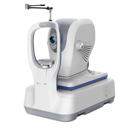
Specifications
VASCAN™ Essential
| Scanning volume/area |
3mm x 3mm 256 x 256 A-scans
12mm x 8 mm 540 x 360 A-scans |
| Algorithm |
C-OMAG |
| Segmentation options |
Encoded, Vitreousretina Intrerface(VRI), Superfcial retina, Deepfcial retinal, Avascular, Choriocapillaris, Choriod |
| Quantitative analysis |
No |
VASCAN™ Advance
| Scanning volume/area |
3mm x 3mm 256 x 256 A-scans
6mm x 6mm 360 x 360 A-scans
8mm x 8mm 360 x 360 A-scans
12mm x 8 mm 540 x 360 A-scans |
| Algorithm |
C-OMAG |
| Segmentation options |
Encoded, Vitreousretina Intrerface(VRI), Superfcial retina, Deepfcial retinal, Avascular, Choriocapillaris, Choriod, Custom |
| Quantitative analysis |
Yes |